Report / Edited by: Roberta Kline, MD

Dr. Tolga Ozmen is a breast surgical oncologist affiliated with Mass General Hospital and is a faculty at Harvard Medical School. He highlights that the inventor of this novel wearable ultrasound device is Dr. Canan Dagdeviren and her team at the MIT Media Lab. Dr. Dagdeviren, known for her distinguished contributions to science, shares a common research interest with Dr. Ozmen which is advancing breast cancer detection and early diagnosis, which has brought their paths together.
<
The results of our preliminary study are very compelling. In the next phase of our study, we will be exploring the performance of our device in a larger cohort of patients. Our objective is to confirm a baseline of (at least) the same performance as the conventional models – within a much larger number of patients. And if everything goes as planned, I don't see any reason why we can't hit the market in the next 10 years.
 |
| Source: MIT News |
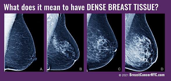
The current standard in breast cancer screening for average risk women includes yearly breast exams and mammograms starting at age 40. This is an effective screening method for decreasing breast cancer mortality in average risk population, backed by numerous published research studies. But we also know that mammography has been shown to be inferior to other imaging modalities in certain cases, including women with dense breast tissue. In the presence of moderate or extreme density of breast tissue, the sensitivity of mammogram decreases significantly, reducing the ability to detect breast cancer. In these women, supplemental imaging modalities in addition to mammography are often indicated.
In some cases, there are findings on the screening that require close follow up. Especially if these women are already identified as being at higher risk, they would benefit from follow up in 3-6 months rather than the standard one year.

We potentially see patients wearing this device as often as every couple of months, enabling us to detect changes early. There are often scenarios where screening mammography and/or ultrasound may show abnormalities that aren’t concerning enough to warrant biopsy but do benefit from close follow-up. Often the patient is requested to come back in 3-6 months. Wearing this device between mammogram visits could offer new data in the form of significant changes in the size or characteristics of that abnormality. Any new images can be generated and reviewed at the central unit, and compared with previous imaging. This interventional imaging allows us to watch the changes on a particular mass or lesion more closely and intervene sooner than their regular screening schedule when warranted.

Breast cancer screening itself doesn't only include mammograms or other imaging. It also includes a patient's self‐awareness, and we recommend women start doing self breast exams by the age 20‐25 and knowing the signs, symptoms, and risk factors that increase (and decrease) breast cancer risk. This expansion of what is considered part of breast cancer screening is very important, and is why we're trying to do as much as we can in order to increase awareness.
Assessing a woman’s risk enables us to further personalize her strategies and screening schedule. High risk patients may have inherited gene mutations, a family history, or both. For these women we typically start screening them with a mammogram or MRI 10 years younger than the first family member with breast cancer. For younger women that start at age 20‐25, we typically will screen with an MRI until after age 30, when we may add mammography as per recommended guidelines.
Reference & Image Sources: https://news.mit.edu/2023/wearable‐ultrasound‐scanner‐breast‐cancer‐0728


"As Breast cancer continues to be the second leading cause of cancer death in women, the battle continues and one of our best weapons in this fight is the innovative use of technology. Screening and diagnostic protocols are a major part of imaging but so is image guidance and monitoring. This technology offers significant promise in the tracking and treatment support on a logistical level. The idea of a wearable imaging monitor gives both the patient and the physician better data as to treatment progress and/or potential malignancy. It uses remote/wifi technology in the best way- by connecting the patient directly to the central monitoring specialist in real-time, eliminating any delays and appointment waiting." - Dr. Robert L. Bard -breast cancer radiologist, NYC


The State of Connecticut passed legislation requiring notification of breast density in 2009, after having passed legislation requiring insurance coverage for ultrasound for dense breasts in 2005. As an ObGyn physician practicing in CT at the time, I remember the discussions with colleagues and patients around this issue although at the time there were no formal efforts to raise awareness or update guidelines from our national specialty organization, the American College of Obstetricians and Gynecologists (ACOG).
PERSPECTIVE: PERSONAL FINDINGS BY A CLINICAL PROFESSIONAL
.jpg)
There is still much to be learned about what causes dense breasts and why women with dense breasts have an increased risk of breast cancer, and our ongoing study is one of many that are seeking to answer these questions at the molecular and genetic level. But the evidence that supplementing mammograms with other imaging modalities can increase the rate of early detection is substantial, and provides us with tools we can use right now to make a difference. [3,4] Despite this progress, there are still significant hurdles in changing the standard of care. A recent experience with my routine breast cancer screening highlighted the ongoing challenges. When I had asked to schedule an ultrasound with my screening mammogram, I was informed that it was not done this way – I could only get a mammogram. After my mammogram, I had to wait to receive my letter in the mail approximately one week later to be able to take any additional steps. The interpretation included a description of breast density and recommended to discuss any additional care with my physician.
 |
| See 2022 Dense Breast Ultrasound Study |
Fortunately all was fine, but had I not been a physician that was fully aware of this issue, I would very likely have had only a mammogram and walked away with a dangerously false sense of security. This experience highlighted for me how much still needed to be done more than 20 years after my first experience. Legislation is only part of the solution. Clinician education and public awareness are the keys to changing how the intention behind these laws gets translated into actual change in health care.
 EPILOGUE: CURRENT STANDARDS VS NEEDS
ACOG still officially does not recommend any further imaging for women with dense breasts on mammogram, despite the significant body of evidence suggesting that mammogram alone is insufficient and adjunctive imaging with ultrasound or MRI increases rate of early detection. [5] The U.S. Preventive Task Force [6] does not recommend routine adjunctive imaging for screening women with dense breasts. This leaves many healthcare practitioners, from ObGyns to other primary care providers, unprepared to discuss this with their patients or provide sound recommendations.
The American College of Radiologists, who also publishes the BIRADS standards for breast cancer screening, acknowledges awareness of breast density detection issues with mammography but stops short of recommending routine adjunctive imaging. Instead, they list ultrasound and MRI as “may be appropriate”. [7] We have enough evidence to know how to better serve women with dense breasts, and we can do better. Now we need to push for better education of all primary health care providers, including ObGyns, and continue to raise awareness for women around current knowledge and best practices.


 WHAT ABOUT IF YOU'RE TOO YOUNG FOR A MAMMOGRAM? I went to my doctor for a lump I felt in my breast and she gave me a response that se...
 FOR IMMEDIATE RELEASE NATIONAL CANCER ADVOCACY GROUP RECOGNIZES BARD DIAGNOSTIC RESEARCH FOR ADVANCEMENTS IN DENSE BREAST IMAGING 8/28/202...
 1/19-2023- The ARE YOU DENSE ADVOCACY committee continued to pursue the public importance of patients receiving their own personal medical i...