Wednesday, November 29, 2023

Cohort Study (Part 2)- Diagnostic Scans of Dense Breast subjects

 REMOTE IMAGING INTERPRETATION FROM THE DIAGNOSTIC INVESTIGATOR
By: Dr. Robert L. Bard

My role in this dense breast cohort study is to review all field imaging scans and to generate pathology reports to identify or confirm breast density and breast health of all the test subjects acquired remotely.  Where Dr. Noelle Cutter scans patients out on the field (in her Molloy University campus and in athletic races), our partnership shows the flexibility of ultrasound screening of breast health, density and disease.  Our focus is in women under 39, and women with dense breasts, which is more common in athletic women and women under 40 who are below the age of screening.  We are also able to prove the ease of use and the safety factor of ultrasound as a non-radiation utility. It captures images in real-time with quantitative accurate in finding cancers or benign cysts in dense or lumpy breast tissue. 

Dr. Cutter approached this project as more of a SURVEY, collecting data in various underserved communities- seeking out breast density in low BMI subjects as well as specific cultural groups.  Her objectives aim to detect breast cancers in women under 39, or women with dense breasts that may not (yet) have the means or the capacity to access routine mammography or even clinical palpation by a physician. 

*** See Dr. Noelle Cutter's Overview on this Phase 1 Study of Dense Breast Survey


SCANNER OF CHOICE: THE TERASON 3200t uSmart Portable Ultrasound 
Upon review of a wide range of portable ultrasound devices, we selected the Terason 3200t (hereinafter shall be referred to as the Terason) due to its proven quality, user-friendly interface and its rugged design.  Its capacity offers high resolution imaging and high quality probes that can capture anatomical scans comparable to hospital grade appliances. Hence, we are able to see not only the breast tissue, but also the skin.  This means that if there's a breast cancer that's infiltrating the skin from below (which a clinician may not be able to see and oftentimes cannot feel) this will show that the cancer is a stage two because it's already broken through the bottom layer of the skin. 

HUMAN RE-INTERVENTION IN IMAGING
While everybody's talking about artificial intelligence, the cancer imaging society continues to rely on seasoned experience of a seasoned diagnostic over-reader. Specifically, if there's a benign tumor that's calcified, it has the same look and effect as a breast cancer that's not calcified because calcium and cancer absorbs the ultrasound frequency the same way- whereby the area behind them is basically black, not white. So it takes an experienced eye to determine if something suspicious is really serious.  As of now, artificial intelligence has not yet reached that level of sophistication in detective work. 

WIFI SUPPORTS THE MEDICAL COLLABORATION PARADIGM
Today’s entire emergency response system is well underway the use of WIFI connected devices in their emergency vehicles. The portable rescue services are completely reliant on WIFI connections, allowing for field responders to receive medical guidance and diagnostic reading support from ‘central command’.  Having access of the supervision of a senior medical professional on the field can save lives where precious seconds count- way before the patient arrives in the emergency room. The ability to have wifi allows the responder and the surgical team to confirm the pathology while receiving expert guidance from anywhere in the world within seconds.

PATIENT 1: STUDY OF BREAST LAYERS
This scan (Patent #1) shows that directly under the white line (A) is a black area that is the epidermis of the skin. The horizontal white band below is the dermis. The resolution of the Terason ultrasound device allows us to see less than half millimeter lesions. Below this is an area (B) that's mixed white and dark. It's got horizontal bands of white and broad swaths of dark fatty tissue, white is breast tissue.  The scan shows normal breast tissue, which is gray.


PATIENT 2: ULTRASOUND DEPTH CAPACITY
To contrast from patient 1, we have band of white tissue (C), which is normal fatty breast tissue- appearing as white. Below C is the semi-circular bands represent the rib cage and more importantly, the surface of the lung. This is useful because should a cancer of the breast metastasize to the lymph nodes or to the lungs, this is where we might see evidence of this during this simple screening examination. The dense breast tissue is the band of white above the semi-circular rib cage echoes. And in the middle of the band of whites, you could see tiny black wormy lines, which represent dilated ducts, which are a part of breast disease or cystic breast disease.


PATIENT 6: CATEGORY 4 DENSITY
Once again, the skin outline (Epidermal) is on the top, which the curvilinear ribs in the middle of the scan, which is a broad white path. And within the middle of this white path are tiny, bright white areas representing micro calcium inside the breast. The fibrous tissue is an inflammatory process which heals with scarring and calcification, which can also block the ducts and dilate the ducts. So here we have an example of a very white dense breast with areas of micro calcification, which are part of the healing process. This would be a CATEGORY 4 breast density on mammography because the tissue is homogeneously white.


PATIENT 7: BENIGN TUMOR
Here's an example of a benign tumor within the breast. Right below the word, right below the B. You have a black area within the white carpet of tissue, and it is sharply demarcated. It has echoes behind it. That is, there's a, a white trail behind the black area showing that it's a degenerated cystic area or a solid benign tumor that's breaking down into cystic regions. So this is an area that we follow, uh, every six months with ultrasound scans. This would show up as a BI-RADS or as a class four density and probably be missed on mammograms. In a prior research project, we did a study showing that the benign tumors like this in dense breasts are missed about 90% of the time by mammography. And of course, since the breast tissue is lumpy, they're often hard to palpate clinically.


PATIENT 8: CATEGORY 1 DENSITY
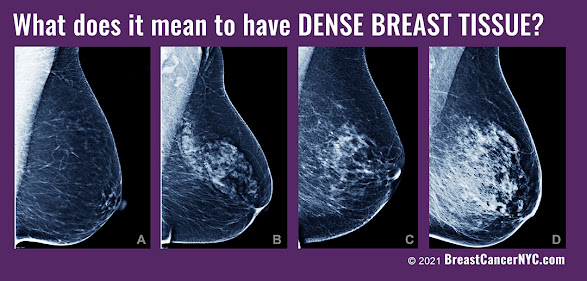
This is an example of a one density mammogram or breast tissue, because this is almost uniformly dark or fatty tissue within the breast.  The linear white lines are prominent because the bulk of the tissue is predominantly fatty


PATIENT 9: CATEGORY 3 WITH MICROCALCIFICATION
This is an example of a class three.  Here, we can see the white tissue interrupted by horizontal areas of dark and white indicating dilated ducts, some of which have micro calcification within them. Once again, we see the chest wall below that. So if there is a cancer, we can see if it's invading not only the skin, but the muscle tissue below that. With this technology, we can find cancers and stage them within minutes.


PATIENT #10:
This is another example of mixed density tissue because you have alternating bands of gray and darker gray. Below that is a very thick pectoral muscle below, which is the white band of the rib cage. So we can see muscle disease as well as the pleural disease (on the lining of the lung).  We can also measure the thickness of a muscle. This may be useful in treating muscle wasting diseases.


FINAL STATEMENT
Breast density is a key factor in early detection because mammograms routinely miss breast cancer in dense breasts, especially in younger and more athletic women. Ultrasound offers a supplemental scan and peace of mind because it finds the pathology and almost instantaneously distinguishes from a benign cyst from a possible cancer - or a definite cancer. Ultrasound even goes further because if there is a definite cancer or something highly suspicious of a cancer, you can look at the other organs such as the glands under the arm to see if it's spread into the lining of the lungs to see if it's invading the pleura or the lymph nodes in the abdomen or the brain. Since we are now armed with expert diagnostic over-readers and remote collaborative imaging options, I urge our gynecologic and obstetric community to take advantage of this affordable non-invasive screening solution. 


Tuesday, November 21, 2023

Cohort Study (Part 1)- Dense Breast Scanning Survey / Molloy University

ACADEMIC PREFLIGHT STUDY OF ULTRASOUND SCREENING APPLICATIONS FOR DENSE BREAST CASES

Transcript by: Noelle Cutter, Ph.D.
Edited by: Roberta Kline, MD and Lennard M. Goetze, Ed.D

We started working with the Terason 3200t Ultrasound (hereinafter shall be referred to as the Terason) in the summer of 2022. The goal of the project was to be able to scan a limited number of younger premenopausal women under the age of 40 who are more likely to have dense breasts based on their status as being endurance based athletes. We have been exclusively using the Terason based on the recommendation of our lead radiologist Dr. Robert Bard.  The Terason became our lab scanner of choice to conduct the screening of 10 individuals in the lab on a voluntary basis to get a readout for their breast density. Endurance athletes are more prone to dense breast tissue, especially athletes under the age of 40. Mammographic density is the strongest risk factor for non-familial breast cancer among women, especially those under the age of 40.   Metastasis, tumor relapse, and resistance to therapy remain the principal causes of death for breast cancer patients and the lack of effective therapies calls for an improved understanding of the molecular mechanisms driving breast cancer progression. Early identification of mammographic density is an essential step in preventative health.


REMOTE COLLABORATIVE TEAM
The Dense Breast paradigm continues to plague our population of the underdiagnosed and underserved women. uniting with public advocates and legislative change makers like the ARE YOU DENSE? FOUNDATION raises significant public awareness in both the clinical and patient communities (see news feature).  Meanwhile, this cohort study received great support from the medical research and diagnostic society whereby scientific leaders like Dr. Robert L. Bard (diagnostic imaging specialist) and Dr. Roberta Kline (epigenetics expert in women's health) helped to spearhead their own respective areas of study to help offer new insight and data in support of the launch of this field research program.


DENSE BREAST CASES
We assembled 10 volunteers for our initial review. These subjects were all selected from the triathlete community who recently either participated in the full distance 140.6 Ironman or the half distance 70.3 Ironman in 2022.  From Molloy University (base 1 of our research), I recruited from our student population who also worked in the lab- as well as from my professional contacts in the endurance world. 

To note, out of our select volunteers, three have been clinically diagnosed with breast cancer within the last five years. I would say that out of the two women who had cancer, both had dense breasts, and both were Ironman athletes under the age of 40 when they were diagnosed. They're both over the age of 40 now. So, they knew that already going into our scanning. I am glad to also report that both cases remain in remission.  Noting these cases, we did not consider using them as a control by any means, only that this is part of the data that we are collecting as additional considerations that relate to our survey for tissue density, as a means of comparison. 

The other eight subjects were young women all under the age of 35- and none of them had any idea about breast density. They've never had any breast screenings in their life besides self-breast examinations- hence, they had no idea about density. Furthermore, they had no idea of how mammographic density is really one of the strongest risk factors for non-familial breast cancer among women and especially among younger women. Undergoing our 'simple' screening session gave them the beginnings of a sense of personal ownership and advocacy about their health.  Perhaps this experience (for them) could lead to proactive steps that they need to take- the next time they visit with either their OBGYN or their general physician. They now have an idea about the types of tests to request to be proactive about their health They also have some clarity about mammographic density in their breasts. 

PORTABLE, HOSPITAL GRADE AND FIELD FRIENDLY
My technicians and I found the Terason to be "super portable" and upon scanning our first few subjects, we immediately understood why Dr. Bard suggested this brand and model. The picture quality and imaging system is very clear and the user interface is very easy to read. For someone who is not experienced or well trained in using an ultrasound, with one or two training sessions I was able to easily use the machine to work with our volunteer subjects. 

An underlying test is the feasibility of using the Terason itself in actual field work. The portability is astounding (to me). This means we can pack it up. I don't even need any sort of a car. I could throw it on my back and get on a bicycle and bring it to a, uh, a destination.  As far as its' performance, my group and I did our own homework about its accuracy and reliability and what we often found was the common statement about it being "battle-tested"- in the EMS world). Targeting the triathlete community, all these features are critical advantages to roadside screening! It's easy to use and even easier to train other research assistants in the lab to use it. And with Dr. Bard (and his expert interpretation) on the receiving end of our scans, that is the ultimate peace of mind and a COMPLETE tele-health scanning paradigm. 


To note, our research concept focused on using the ultrasound because it is completely safe and effective at scanning for (something like) image breast density. Particularly for our research, it's portable in the field, so we can bring it to the race locations. I can bring it into the lab, I can bring it into my office. I can really package it up in a small briefcase sized bag, and it can come with me and travel with me anywhere I need to go. 


REMOTE ULTRASOUND TRAINER
Working with Mike (Terason virtual trainer) was surprisingly easy, and he thoroughly guided me over the phone and via the device itself which is also a computer. He gave us such a comprehensive tour on all of the different probes but of course, we were focused on the one (L20) probe that was suited for of screening of breast density.  We found the device itself to cover the most common demands of a hospital-grade or field-grade ultrasound including the ability to capture measurements of critical data like blood flow and density. Recalling my initial training session with Dr. Bard (at his office) aligned with everything because as a field/remote ultrasound tech, I am literally capturing all images for him (our in-house radiologist).  This Terason model truly enables this remote scanning paradigm between the field screening tech in communication with the central radiologist- making for a truly ideal and real time professional collaboration!   (To be continued below)

Epigenetic Research Notes: Profiling the Dense Breast Paradigm
A Clinical Report by: Dr. Roberta Kline


As an Ob-Gyn physician and genomics specialist, I have spent the better part of 10 years translating research in the genomic and gene expression areas into clinically usable information for healthcare professionals. One of the biggest challenges we face when connecting research with patient care is the long delay in the translation process and dissemination of the information. It often takes 10 to 20 years for information (validated findings) that comes out of research to be applied in clinical practice. These delays result in many lost opportunities to provide better care for our patients. This is one of the reasons why I'm really passionate about accelerating this process and making it easier for clinicians and their patients to take advantage of cutting-edge information and new technologies. 

VITAL RESEARCH IN SUPPORT OF EARLY DETECTION
I believe that because our focus was on women with dense breasts and the fact that this issue is now showing escalating rates, the urgency for this project also aligns with an increased likelihood of developing cancer within their lifetime. We also know that women, especially pre-menopausal women, are often not sent for imaging unless they have a family history. But we know only about five to 10% of breast cancers occur because of an inherited or genetic variant. So the likelihood that we're missing a lot of women (especially young women) who may have an early stage of breast cancer is the big concern. 

This project was developed out of the need for a safe, effective, and affordable screening alternative. We wanted to explore and possibly attest to the higher benefits of this modern (and field-friendly) alternative vs. the existing "gold standards" which are far more inaccessible, expensive and potentially invasive- as far contrast or radiation. I appreciate my understanding of ultrasound as a fully safe and effective imaging modality - and one that we can use and easily can be accessed in any physician's office or anywhere else. 

After our dedicated training course, the roll-out to being a stage 1 ultrasound technician was very comfortable and truly supportive of our goals about ultrasound as the path to the future and "the better way".

As far as goals, our main objectives are (of course) advocacy and to identify women younger using a more safe and effective screening method. Another goal that we found of great interest in enabling ACCESS to women have dense breasts; we wanted to explore ways that we can get them screened sooner. We can have them checked out to make sure that there are no early cancers that are being missed through other tools and techniques that we can use. In addition to this, we can start looking at how density correlates to cancer. Our early studies indicate that many of the pathways enriched in patients with higher mammographic density are targetable, raising the possibility of developing prevention strategies for mitigating density-associated breast cancer risk. We hope to further elucidate those molecular changes and those genetic signatures that may be common in women with dense breasts. Our overall goal is to increase survivability and offer targetable outcomes for the patient. 


PROGRESSIVE CLINICAL MODALITIES IN ADDRESSING ARTHRITIS SYMPTOMS

By: Robert L. Bard, MD / Lennard Goetze, Ed.D   [PDF DOWNLOAD] While arthritis starts as bone and cartilage degeneration, oftentimes the fi...