Monday, December 18, 2023

EFFECTS OF PULSED BIOENERGY ON PROSTATE HYPERPLASIA (ENLARGEMENT)- 10/2023


ABSTRACT
On average, the prostate is about the size of a walnut. It grows to roughly the size of a Ping-Pong ball, weighing 30 to 35 grams. Within a major portion of adult males, the prostate continues to grow even further up to the size of a tennis ball or larger. The normal prostate gland has measures 3 × 3 × 5 cm approximately or a volume of 25 ml. [1,2] According to statistical reports by Yale Medicine about 50% of men between the ages of 51 and 60 have Benign Prostatic Hyperplasia (BPH) or enlarged prostate, and that number jumps to 70% among men aged 60 to 69 and around 80% of men over 70 years of age [3].  

Complications of BPH includes acute urinary retention, chronic, or long lasting urinary retention, blood in the urine, urinary tract infections (UTIs), bladder & kidney damage or bladder stones.[4]  The most common solution (thus far) to address this condition is medicinally with Alpha blockers.  This is recognized as the recommended first-line treatment for men with mild to moderate symptoms. It is noted that alpha blockers carry a variety of side effects including dizziness and low blood pressure.[5] There are also invasive solutions to enlarged prostate including laser therapy, microwave heat, or prostate tissue compression. Partial prostate removal and full removal are more invasive but may be necessary for extremely large prostate glands.  

As the medical community is (now) pursuing non-invasive alternatives for the many health conditions, addressing hyperplasia continues to be a significant interest in the medical community.  Bard Diagnostics and Wellness Now, under a joint study under the AngioFoundation(501c3) is conducting a preflight study of the effects of Pulsed Bioenergy Therapeutic innovations.  This Image-Guided program employs the use of PEMF (Pulsed Electromagnetic Frequency) technology, or neuromagnetic stimulation to address symptoms of enlarged prostate.

EFFICACY REVIEW:
Since March of 2022, Dr. Robert Bard initiated a multi-pathology study of PEMF treatments.  This specific study is a clinical evaluation of the PEMF treatments of four (4) volunteers @ the Bard Diagnostic facility at 121 E. 60th St. NYC. In pursuit of the many reports of Electromagnetic Stimulation providing significant success in inflammation reduction, applying the effects of the cellular regenerative properties of neuromagnetic field directly on a pre-confirmed enlarged prostate gland dictates the same logical path of size reduction in an enlarged prostate.

Supervised exposure was provided by Dr. Bard and his research team through the use of real-time 3D Doppler Ultrasound and other non-invasive imaging devices.  The initial (Image Guided) treatment was conducted @ the NYC clinic where Dr. Bard established base line studies and regular (bi-weekly) PEMF treatments and ultrasound scans under periodical comparative imaging.  In addition, each volunteer took home a personal PEMF device for use twice a day at a given power setting and dosage duration. This preflight study was a one month (30 days) micro-review of the PEMF’s function and a logging of the volunteer’s exposure response via imaging.  Dr. Bard recorded progress within the time allotted- collected quantifiable data about the state of the volunteer’s prostate size and health. Progress verified by a reduction in size, blood velocity (flow) within the immediate prostate area as well as the elasticity or firmness of the prostate tissue.  This information was collected by the diagnostic imaging technologies selected for this study at Dr. Bard’s scanning devices. 


 REFERENCES

(1) verage size prostate: https://utswmed.org/medblog/what-we-know-about-your-prostate/#:~:text=On%20average%2C%20the%20prostate%20is%20about%20the%20size%20of%20a%20walnut.&text=It%20grows%20to%20roughly%20the,weighing%2030%20to%2035%20grams.&text=In%20many%20men%2C%20it%20continues,a%20tennis%20ball%20or%20larger. (2) Ultrasound of the prostate – PMC : https://www.ncbi.nlm.nih.gov/pmc/articles/PMC2842183/ (3) Enlarged Prostate (Benign Prostatic Hyperplasia)  https://www.yalemedicine.org/conditions/enlarged-prostate-benign-prostatic-hyperplasia-bph (4) Prostate Enlargement (Benign Prostatic Hyperplasia): https://www.niddk.nih.gov/health-information/urologic-diseases/prostate-problems/prostate-enlargement-benign-prostatic-hyperplasia (5) Alpha Blockers for the Treatment of Benign Prostatic Hyperplasia - https://www.ncbi.nlm.nih.gov/pmc/articles/PMC2213889/ (6) Prostate resection - minimally invasive - https://medlineplus.gov/ency/article/007415.htm (7) "Enlarged Prostate" - PennMedicine.org https://www.pennmedicine.org/for-patients-and-visitors/patient-information/conditions-treated-a-to-z/enlarged-prostate# (8) "Effect of Pulsed Electromagnetic Field Therapy on Prostate Volume and Vascularity in the Treatment of Benign Prostatic Hyperplasia: A Pilot Study in a Canine Model" | National Institutes of Health.- PMID: 24913937- Raffaella Leoci,* Giulio Aiudi, Fabio Silvestre, et al.  https://www.ncbi.nlm.nih.gov/pmc/articles/PMC4145661/ (9) "Therapeutic use of pulsed electromagnetic field therapy reduces prostate volume and lower urinary tract symptoms in benign prostatic hyperplasia"- PMID: 32090492 |  Marta Tenuta, Maria G Tarsitano, Paola Mazzotta et al. https://pubmed.ncbi.nlm.nih.gov/32090492/ (10) "Ultrasound & The Prostate": RadiologyInfo.org |  https://www.radiologyinfo.org/en/info/us- prostate#:~:text=Ultrasound%20of%20the%20prostate%20uses,whether%20the%20gland%20is%20enlarged. (11) "Prostate cancer ultrasound treatment as effective as surgery or radiotherapy", by Kate Wighton (04 July 2018). Imperial College London. https://www.imperial.ac.uk/news/187086/prostate-cancer-ultrasound-treatment-effective-surgery/ (12) "Mechanisms and therapeutic effectiveness of pulsed electromagnetic field therapy in oncology"- 2016 Nov 5, | PMID: 27748048 | Maria VadalĂ , Julio Cesar Morales‐Medina, Et. Al. - https://www.ncbi.nlm.nih.gov/pmc/articles/PMC5119968/


Excerpt from "COUSIN SAL'S PROSTATE HEALTH MISSION"- (source: www.cousinsal.org)

INTRODUCTION: By "Cousin" Sal Banchitta
I've had an incredible 30+ year career in the NY Fire Department, what so many of considered to be the best job in the world. There is no other profession that even comes close to the rewards of being a city firefighter. We were the first and last line of defense to protect this great city from any catastrophe and aligned with a special family of the most unique and remarkable men and women is truly the ultimate blessing.

Many of us will back me on this; there is no such thing as a RETIRED FIREFIGHTER. The very spirit to make a difference as a first responder is a calling that stays with all of us until our last day. We are 'built' to stand watch for any threats to our community and be at the ready to do what needs to be done to restore safety and normalcy to the lives of our neighbors. One of many such examples is 9/11- where so many old-timers and active responders from all over the country raced to New York City at a moment's notice to unite with our fellow rescuers. Seeing our city in peril was an instant call to run to the scene without any hesitation and stand next to our fellow rescuers to work and sacrifice everything all over again.

So when it comes to OCCUPATIONAL INJURIES, EXPOSURES AND CANCERS, I co-captained the 'GET CHECKED NOW!" Project with NYCRA because staying proactive is a must-do! This includes staying on top of some of the major concerns in retirement like PROSTATE HEALTH.  When I heard about this PEMF study, I was the first to join as a test case. I proudly announce this (instead of conceal my identity as with most research projects). I wanted the world to know that exploring alternative therapies greatly shaped the world of health and longevity and that non-invasive monitoring is a big part of what Dr. Bard calls "IMAGE GUIDED TREATMENTS". Whomever sees my articles about this tech review and all others that I am involved in, I hope to project the message of maintaining current awareness about what's available out there- as part of staying on top of your health!


EX-FIREFIGHTER JOINS HEATLH TECH RESEARCH: ADDRESSING ENLARGED PROSTATE

NYCRA's own "COUSIN SAL" BANCHITTA, Retired FDNY FF and Cancer Prevention Advocate for the F.A.C.E.S. (Firefighters Against Cancers and Exposures) "Get Checked NOW!" program often partners with Dr. Robert Bard's clinical research team to explore the latest in diagnostic and therapeutic health innovations.

In August of 2023, Sal joined a group of four men over 50 on an exploratory and clinically monitored study applying PEMF (Pulsed Electromagnetic Frequency) to address BPH (Benign prostatic hyperplasia) or enlarged prostate gland. According to Yale Medicine, "about 50% of men between the ages of 51 and 60 have BPH, and that number jumps to 70% among men aged 60 to 69 and around 80% of men over 70 years of age".[1]

This exploratory concept was under a collaboration between Dr. Robert Bard (seasoned diagnostic imaging specialist) and a team of research coordinators from ICRS (Integrative Cancer Resource Society). In expanded technical collaboration with Mr. Patrick Ziemer (AuraWell ness PEMF), this panel launched an exploratory Pilot concept of employing a non-invasive alternative solution to reduce prostate size as part of improving men's overall health. "Historically, we have observed the growing success of pulsed bioenergy frequency on a wide variety of physiological disorders... joining the strength of Dr. Bard's imaging capacity to monitor and validate real-time treatment progress, we have united to launch this micro study... that may someday change the face of how prostate disorders may be managed", states Dr. Noelle Cutter, ICRS Senior Medical Editor & clinical research specialist.

Use of ultrasound imaging has been Dr. Bard's "scanner of choice" when it comes to analyzing and diagnosing a wide array of complex health disorders, from cancer tumors to inflammatory disorders. As a beta tester for developers of ultrasound feature upgrades, Dr. Bard has published an expanded set of findings in support of PEMF regenerative benefits.  His latest pilot study provided quantitative evidence about PEMF-induced micro-tissue and vascular reactions, thanks to his creative use of echocardiography. "Over the years, I have gained significant confidence in Pulsed Bioenergy therapeutic innovations", states Dr. Bard. "...through strategic imaging, we can visibly and quantifiably identify the smallest veins dilating and the contractility of the tissue being energized- appearing as pulse vibrations in the form of the undulating surface line of the muscle. This is how we can illustrate the path of ENERGY MEDICINE in real-time action".


PILOT STUDY ON THE ROAD
Sal Banchitta, one of four volunteers took home the Nova-HD (by AuraWell PEMF) from the Bard Diagnostic Imaging center in NYC. All case studies are instructed to use the PEMF device as directed- applying electromagnetic exposure via the provided PEMF coil for an est. 20 minutes 2x a day. "Aside from the clicking sound of the power source device, what I learned about PEMF is that it's widely known to be painless and has no known side effects", says Mr. Banchitta. "I also learned this technology to offer so much by ways of pain relief and cell regenerative therapy".

Unlike the other participants of the study, Sal was scheduled to go on a coastal road trip for business immediately after his PEMF consult. The clinical panel found this to be an ideal situation to conduct the test from the drivers seat.  "Adult Americans spend a major portion of their lives behind the wheel... proving how to integrate PEMF treatments while driving could be a great way to optimize use of one's travel time", stated Patrick Ziemer.

TREATMENT "ON THE GO"- By Cousin Sal
"Because I had a limited amount of time and a tight travel schedule, I chose to take advantage of the situation by bringing the PEMF into my truck- and it was easy! I installed an a 110 AC outlet to plug in the device. Sitting on the PEMF coil is the same here as it is in my recliner chair at home. The coil was soft and absolutely comfortable and it wasn't an impediment to my driving at all. I used it for about an hour each day and I had a lot of hours to kill." 


EPILOGUE: SAL'S HEALTH JOURNEY
"I have always been a proponent of early detection- especially when it comes to prostate cancer. Four years ago, my annual retirement exam started with a blood test. My primary found my PSA was slightly elevated, so I was then sent to the urologist as standard operating procedure.  Before you know it, I received a report of a "slightly" enlarged prostate which put me into what felt like an automated track to getting a biopsy.  Getting a biopsy without an MRI or any type of imaging made the needle work a complete and painful shot in the dark. Getting poked 12+ times in what felt like complete guesswork was terrible experience. In the end, we found that the biopsy could have been avoided if the PSA reading gave more information, and a more accurate assessment. As a member of a family predisposed to cancer, the stress of waiting for the biopsy, then actually undergoing the procedure thinking I MAY have have cancer or not was completely an unfair ordeal to put anyone through. Meanwhile, this is what launched my journey to support prostate health- including why I am now driving with a PEMF coil on my drivers seat!"


1) https://www.yalemedicine.org/conditions/enlarged-prostate-benign-prostatic-hyperplasia-bph#:~:text=But%20because%20enlarged%20prostate%2C%20or,over%2070%20years%20of%20age.


SPECIAL THANKS
The news feature proudly gives thanks to the AOPP (Association of PEMF Professionals) and Patrick Ziemer of Magnawave Inc. and Aura Wellness PEMF for the technical information provided for interviews and unending support to support our clinical study and educational programs about PEMF technology for chronic disorders and supportive testimonials in alternative therapeutics.   


(Part 2) IMAGE GUIDED APPROACH TO THE TREATMENT OF HYPERPLASIA  By: Dr. Robert L Bard

Procedurally, the best way to study an enlarged hyperplastic prostate gland is as a low grade inflammatory process, similar to women with fibrocystic breasts considered to be breast inflammation. This does not need to be treated with antibiotics since this inflammation is often aligned with a chronic disease and requires a long-term treatment protocol. One example of this therapy is using a pulse electromagnetic applicator (PEMF) which can be applied over the groin area or under the pelvis (even in the car seat). For treatment applications, this protocol is recognized to be safe, painless, comfortable, and user-friendly on a long-term basis.  Reducing low grade inflammation can be managed with relatively low energy treatment over an extended yet controlled period of time.  Daily application of energy therapy inducers to the prostate and bladder area has been proven to reduce bladder urge, the need to urinate and improve the urine flow out of the enlarged prostate. It also decreases the chance of incontinence because the bladder that is more easily emptied is less likely to overflow in an uncontrolled manner.  (CLICK to see complete article)


SALSCAN 2.0: Cousin Sal Goes WHOLE-BODY MRI
8/8/2023- Ret. FDNY Sal Banchitta signs in to experience his Whole Body MRI from Prenuvo, NYC- a state of the art imaging company founded out of Vancouver, Canada. As one of the founding cancer awareness producers of NYCRA (NY Cancer Resource Alliance0 and F.A.C.E.S. (Firefighters Against Cancers & Exposures), Mr. Banchitta gladly accepted the invitation to receive this complete body scan as part of his pursuits of a base line for future scans and a preventative measure against cancer. "My mission alongside my fellow advocates is to learn about the latest modalities in diagnostics and treatment to bring awareness about cancer resources to the rescue community", says Sal. Prenuvo’s cutting-edge whole-body scans are based on 10+ years of clinical work while curating the biggest data set of whole-body MRI scans in the world. Unlike conventional MRIs, which take hours and often involve contrast injections, Prenuvo scans for 500+ conditions, including most solid tumors which can be detected as early as stage 1, in addition to aneurysms, cysts, and more – all without radiation, in under an hour. (See full article)


Copyright Notice: The materials provided on this website/web-based article are copyrighted and the intellectual property of the publishers/producers (The NY Cancer Resource Alliance/IntermediaWorx inc. and The AngioFoundation). It is provided publicly strictly for informational purposes within non-commercial use and not for purposes of resale, distribution, public display or performance. Unless otherwise indicated on this web based page, sharing, re-posting, re-publishing of this work is strictly prohibited without due permission from the publishers.  Also, certain content may be licensed from third-parties. The licenses for some of this Content may contain additional terms. When such Content licenses contain additional terms, we will make these terms available to you on those pages (which his incorporated herein by reference).The publishers/producers of this site and its contents such as videos, graphics, text, and other materials published are not intended to be a substitute for professional medical advice, diagnosis, or treatment. For any questions you may have regarding a medical condition, please always seek the advice of your physician or a qualified health provider. Do not postpone or disregard any professional medical advice over something you may have seen or read on this website. If you think you may have a medical emergency, call your doctor or 9-1-1 immediately.  This website does not support, endorse or recommend any specific products, tests, physicians, procedures, treatment opinions or other information that may be mentioned on this site. Referencing any content or information seen or published in this website or shared by other visitors of this website is solely at your own risk. The publishers/producers of this Internet web site reserves the right, at its sole discretion, to modify, disable access to, or discontinue, temporarily or permanently, all or any part of this Internet web site or any information contained thereon without liability or notice to you.

Disclaimer: The information (including, but not limited to text, graphics, images and other material) contained in this article is for informational purposes only. No material on this site is intended to be a substitute for professional medical advice or scientific claims. Furthermore, any/all contributors (both medical and non-medical) featured in this article are presenting only ANECDOTAL findings pertaining to the effects and performance of the products/technologies being reviewed - and are not offering clinical data or medical recommendations in any way. Always seek the advice of your physician or other qualified health care provider with any questions you may have regarding a medical condition or treatment and before undertaking a new health care regimen, never disregard professional medical advice or delay in seeking it because of something you read on this  article or blogsite.

Wednesday, November 29, 2023

Cohort Study (Part 2)- Diagnostic Scans of Dense Breast subjects

 REMOTE IMAGING INTERPRETATION FROM THE DIAGNOSTIC INVESTIGATOR
By: Dr. Robert L. Bard

My role in this dense breast cohort study is to review all field imaging scans and to generate pathology reports to identify or confirm breast density and breast health of all the test subjects acquired remotely.  Where Dr. Noelle Cutter scans patients out on the field (in her Molloy University campus and in athletic races), our partnership shows the flexibility of ultrasound screening of breast health, density and disease.  Our focus is in women under 39, and women with dense breasts, which is more common in athletic women and women under 40 who are below the age of screening.  We are also able to prove the ease of use and the safety factor of ultrasound as a non-radiation utility. It captures images in real-time with quantitative accurate in finding cancers or benign cysts in dense or lumpy breast tissue. 

Dr. Cutter approached this project as more of a SURVEY, collecting data in various underserved communities- seeking out breast density in low BMI subjects as well as specific cultural groups.  Her objectives aim to detect breast cancers in women under 39, or women with dense breasts that may not (yet) have the means or the capacity to access routine mammography or even clinical palpation by a physician. 

*** See Dr. Noelle Cutter's Overview on this Phase 1 Study of Dense Breast Survey


SCANNER OF CHOICE: THE TERASON 3200t uSmart Portable Ultrasound 
Upon review of a wide range of portable ultrasound devices, we selected the Terason 3200t (hereinafter shall be referred to as the Terason) due to its proven quality, user-friendly interface and its rugged design.  Its capacity offers high resolution imaging and high quality probes that can capture anatomical scans comparable to hospital grade appliances. Hence, we are able to see not only the breast tissue, but also the skin.  This means that if there's a breast cancer that's infiltrating the skin from below (which a clinician may not be able to see and oftentimes cannot feel) this will show that the cancer is a stage two because it's already broken through the bottom layer of the skin. 

HUMAN RE-INTERVENTION IN IMAGING
While everybody's talking about artificial intelligence, the cancer imaging society continues to rely on seasoned experience of a seasoned diagnostic over-reader. Specifically, if there's a benign tumor that's calcified, it has the same look and effect as a breast cancer that's not calcified because calcium and cancer absorbs the ultrasound frequency the same way- whereby the area behind them is basically black, not white. So it takes an experienced eye to determine if something suspicious is really serious.  As of now, artificial intelligence has not yet reached that level of sophistication in detective work. 

WIFI SUPPORTS THE MEDICAL COLLABORATION PARADIGM
Today’s entire emergency response system is well underway the use of WIFI connected devices in their emergency vehicles. The portable rescue services are completely reliant on WIFI connections, allowing for field responders to receive medical guidance and diagnostic reading support from ‘central command’.  Having access of the supervision of a senior medical professional on the field can save lives where precious seconds count- way before the patient arrives in the emergency room. The ability to have wifi allows the responder and the surgical team to confirm the pathology while receiving expert guidance from anywhere in the world within seconds.

PATIENT 1: STUDY OF BREAST LAYERS
This scan (Patent #1) shows that directly under the white line (A) is a black area that is the epidermis of the skin. The horizontal white band below is the dermis. The resolution of the Terason ultrasound device allows us to see less than half millimeter lesions. Below this is an area (B) that's mixed white and dark. It's got horizontal bands of white and broad swaths of dark fatty tissue, white is breast tissue.  The scan shows normal breast tissue, which is gray.


PATIENT 2: ULTRASOUND DEPTH CAPACITY
To contrast from patient 1, we have band of white tissue (C), which is normal fatty breast tissue- appearing as white. Below C is the semi-circular bands represent the rib cage and more importantly, the surface of the lung. This is useful because should a cancer of the breast metastasize to the lymph nodes or to the lungs, this is where we might see evidence of this during this simple screening examination. The dense breast tissue is the band of white above the semi-circular rib cage echoes. And in the middle of the band of whites, you could see tiny black wormy lines, which represent dilated ducts, which are a part of breast disease or cystic breast disease.


PATIENT 6: CATEGORY 4 DENSITY
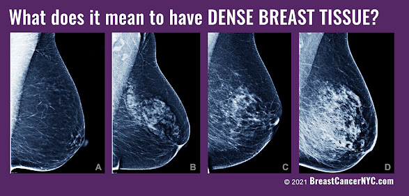
Once again, the skin outline (Epidermal) is on the top, which the curvilinear ribs in the middle of the scan, which is a broad white path. And within the middle of this white path are tiny, bright white areas representing micro calcium inside the breast. The fibrous tissue is an inflammatory process which heals with scarring and calcification, which can also block the ducts and dilate the ducts. So here we have an example of a very white dense breast with areas of micro calcification, which are part of the healing process. This would be a CATEGORY 4 breast density on mammography because the tissue is homogeneously white.


PATIENT 7: BENIGN TUMOR
Here's an example of a benign tumor within the breast. Right below the word, right below the B. You have a black area within the white carpet of tissue, and it is sharply demarcated. It has echoes behind it. That is, there's a, a white trail behind the black area showing that it's a degenerated cystic area or a solid benign tumor that's breaking down into cystic regions. So this is an area that we follow, uh, every six months with ultrasound scans. This would show up as a BI-RADS or as a class four density and probably be missed on mammograms. In a prior research project, we did a study showing that the benign tumors like this in dense breasts are missed about 90% of the time by mammography. And of course, since the breast tissue is lumpy, they're often hard to palpate clinically.


PATIENT 8: CATEGORY 1 DENSITY
This is an example of a one density mammogram or breast tissue, because this is almost uniformly dark or fatty tissue within the breast.  The linear white lines are prominent because the bulk of the tissue is predominantly fatty


PATIENT 9: CATEGORY 3 WITH MICROCALCIFICATION
This is an example of a class three.  Here, we can see the white tissue interrupted by horizontal areas of dark and white indicating dilated ducts, some of which have micro calcification within them. Once again, we see the chest wall below that. So if there is a cancer, we can see if it's invading not only the skin, but the muscle tissue below that. With this technology, we can find cancers and stage them within minutes.


PATIENT #10:
This is another example of mixed density tissue because you have alternating bands of gray and darker gray. Below that is a very thick pectoral muscle below, which is the white band of the rib cage. So we can see muscle disease as well as the pleural disease (on the lining of the lung).  We can also measure the thickness of a muscle. This may be useful in treating muscle wasting diseases.


FINAL STATEMENT
Breast density is a key factor in early detection because mammograms routinely miss breast cancer in dense breasts, especially in younger and more athletic women. Ultrasound offers a supplemental scan and peace of mind because it finds the pathology and almost instantaneously distinguishes from a benign cyst from a possible cancer - or a definite cancer. Ultrasound even goes further because if there is a definite cancer or something highly suspicious of a cancer, you can look at the other organs such as the glands under the arm to see if it's spread into the lining of the lungs to see if it's invading the pleura or the lymph nodes in the abdomen or the brain. Since we are now armed with expert diagnostic over-readers and remote collaborative imaging options, I urge our gynecologic and obstetric community to take advantage of this affordable non-invasive screening solution. 


Tuesday, November 21, 2023

Cohort Study (Part 1)- Dense Breast Scanning Survey / Molloy University

ACADEMIC PREFLIGHT STUDY OF ULTRASOUND SCREENING APPLICATIONS FOR DENSE BREAST CASES

Transcript by: Noelle Cutter, Ph.D.
Edited by: Roberta Kline, MD and Lennard M. Goetze, Ed.D

We started working with the Terason 3200t Ultrasound (hereinafter shall be referred to as the Terason) in the summer of 2022. The goal of the project was to be able to scan a limited number of younger premenopausal women under the age of 40 who are more likely to have dense breasts based on their status as being endurance based athletes. We have been exclusively using the Terason based on the recommendation of our lead radiologist Dr. Robert Bard.  The Terason became our lab scanner of choice to conduct the screening of 10 individuals in the lab on a voluntary basis to get a readout for their breast density. Endurance athletes are more prone to dense breast tissue, especially athletes under the age of 40. Mammographic density is the strongest risk factor for non-familial breast cancer among women, especially those under the age of 40.   Metastasis, tumor relapse, and resistance to therapy remain the principal causes of death for breast cancer patients and the lack of effective therapies calls for an improved understanding of the molecular mechanisms driving breast cancer progression. Early identification of mammographic density is an essential step in preventative health.


REMOTE COLLABORATIVE TEAM
The Dense Breast paradigm continues to plague our population of the underdiagnosed and underserved women. uniting with public advocates and legislative change makers like the ARE YOU DENSE? FOUNDATION raises significant public awareness in both the clinical and patient communities (see news feature).  Meanwhile, this cohort study received great support from the medical research and diagnostic society whereby scientific leaders like Dr. Robert L. Bard (diagnostic imaging specialist) and Dr. Roberta Kline (epigenetics expert in women's health) helped to spearhead their own respective areas of study to help offer new insight and data in support of the launch of this field research program.


DENSE BREAST CASES
We assembled 10 volunteers for our initial review. These subjects were all selected from the triathlete community who recently either participated in the full distance 140.6 Ironman or the half distance 70.3 Ironman in 2022.  From Molloy University (base 1 of our research), I recruited from our student population who also worked in the lab- as well as from my professional contacts in the endurance world. 

To note, out of our select volunteers, three have been clinically diagnosed with breast cancer within the last five years. I would say that out of the two women who had cancer, both had dense breasts, and both were Ironman athletes under the age of 40 when they were diagnosed. They're both over the age of 40 now. So, they knew that already going into our scanning. I am glad to also report that both cases remain in remission.  Noting these cases, we did not consider using them as a control by any means, only that this is part of the data that we are collecting as additional considerations that relate to our survey for tissue density, as a means of comparison. 

The other eight subjects were young women all under the age of 35- and none of them had any idea about breast density. They've never had any breast screenings in their life besides self-breast examinations- hence, they had no idea about density. Furthermore, they had no idea of how mammographic density is really one of the strongest risk factors for non-familial breast cancer among women and especially among younger women. Undergoing our 'simple' screening session gave them the beginnings of a sense of personal ownership and advocacy about their health.  Perhaps this experience (for them) could lead to proactive steps that they need to take- the next time they visit with either their OBGYN or their general physician. They now have an idea about the types of tests to request to be proactive about their health They also have some clarity about mammographic density in their breasts. 

PORTABLE, HOSPITAL GRADE AND FIELD FRIENDLY
My technicians and I found the Terason to be "super portable" and upon scanning our first few subjects, we immediately understood why Dr. Bard suggested this brand and model. The picture quality and imaging system is very clear and the user interface is very easy to read. For someone who is not experienced or well trained in using an ultrasound, with one or two training sessions I was able to easily use the machine to work with our volunteer subjects. 

An underlying test is the feasibility of using the Terason itself in actual field work. The portability is astounding (to me). This means we can pack it up. I don't even need any sort of a car. I could throw it on my back and get on a bicycle and bring it to a, uh, a destination.  As far as its' performance, my group and I did our own homework about its accuracy and reliability and what we often found was the common statement about it being "battle-tested"- in the EMS world). Targeting the triathlete community, all these features are critical advantages to roadside screening! It's easy to use and even easier to train other research assistants in the lab to use it. And with Dr. Bard (and his expert interpretation) on the receiving end of our scans, that is the ultimate peace of mind and a COMPLETE tele-health scanning paradigm. 


To note, our research concept focused on using the ultrasound because it is completely safe and effective at scanning for (something like) image breast density. Particularly for our research, it's portable in the field, so we can bring it to the race locations. I can bring it into the lab, I can bring it into my office. I can really package it up in a small briefcase sized bag, and it can come with me and travel with me anywhere I need to go. 


REMOTE ULTRASOUND TRAINER
Working with Mike (Terason virtual trainer) was surprisingly easy, and he thoroughly guided me over the phone and via the device itself which is also a computer. He gave us such a comprehensive tour on all of the different probes but of course, we were focused on the one (L20) probe that was suited for of screening of breast density.  We found the device itself to cover the most common demands of a hospital-grade or field-grade ultrasound including the ability to capture measurements of critical data like blood flow and density. Recalling my initial training session with Dr. Bard (at his office) aligned with everything because as a field/remote ultrasound tech, I am literally capturing all images for him (our in-house radiologist).  This Terason model truly enables this remote scanning paradigm between the field screening tech in communication with the central radiologist- making for a truly ideal and real time professional collaboration!   (To be continued below)

Epigenetic Research Notes: Profiling the Dense Breast Paradigm
A Clinical Report by: Dr. Roberta Kline


As an Ob-Gyn physician and genomics specialist, I have spent the better part of 10 years translating research in the genomic and gene expression areas into clinically usable information for healthcare professionals. One of the biggest challenges we face when connecting research with patient care is the long delay in the translation process and dissemination of the information. It often takes 10 to 20 years for information (validated findings) that comes out of research to be applied in clinical practice. These delays result in many lost opportunities to provide better care for our patients. This is one of the reasons why I'm really passionate about accelerating this process and making it easier for clinicians and their patients to take advantage of cutting-edge information and new technologies. 

VITAL RESEARCH IN SUPPORT OF EARLY DETECTION
I believe that because our focus was on women with dense breasts and the fact that this issue is now showing escalating rates, the urgency for this project also aligns with an increased likelihood of developing cancer within their lifetime. We also know that women, especially pre-menopausal women, are often not sent for imaging unless they have a family history. But we know only about five to 10% of breast cancers occur because of an inherited or genetic variant. So the likelihood that we're missing a lot of women (especially young women) who may have an early stage of breast cancer is the big concern. 

This project was developed out of the need for a safe, effective, and affordable screening alternative. We wanted to explore and possibly attest to the higher benefits of this modern (and field-friendly) alternative vs. the existing "gold standards" which are far more inaccessible, expensive and potentially invasive- as far contrast or radiation. I appreciate my understanding of ultrasound as a fully safe and effective imaging modality - and one that we can use and easily can be accessed in any physician's office or anywhere else. 

After our dedicated training course, the roll-out to being a stage 1 ultrasound technician was very comfortable and truly supportive of our goals about ultrasound as the path to the future and "the better way".

As far as goals, our main objectives are (of course) advocacy and to identify women younger using a more safe and effective screening method. Another goal that we found of great interest in enabling ACCESS to women have dense breasts; we wanted to explore ways that we can get them screened sooner. We can have them checked out to make sure that there are no early cancers that are being missed through other tools and techniques that we can use. In addition to this, we can start looking at how density correlates to cancer. Our early studies indicate that many of the pathways enriched in patients with higher mammographic density are targetable, raising the possibility of developing prevention strategies for mitigating density-associated breast cancer risk. We hope to further elucidate those molecular changes and those genetic signatures that may be common in women with dense breasts. Our overall goal is to increase survivability and offer targetable outcomes for the patient. 


Tuesday, September 26, 2023

HealthTech Report #224: Interventional Monitoring of Muscle Reaction to PEMF Induction

Introduction: 
Test conducted on August 18, 2023. In pursuit of scientific data through independent clinical  testing, HealthTech Reporter collaborated with the Integrative Health Research Center (NYC) to conduct this private single case study and efficacy monitoring of biometric response of PEMF  frequency waves.  

Dr. Robert Bard, chief investigator designed this case study with  high resolution 3D RESONA 7 Ultrasound System by MINDRAY, NA. (complete technical info below).

The use of  of the Aurawell PEMF (AuraWave model) was applied during this test. ONE clinical participant was pre-qualified, then exposed to PEMF treatment to the QUADRICEPS muscle area for a given period of time and under a specific frequency.  The clinical imaging specialist employed various applications of a medical-grade ultrasound to monitor any visible physical reactions or movements during the time of the bioenergy treatment exposure.


PHASE 1: PREFLIGHT EXPLORATORY MICRO-TEST
Prior to launching a comprehensive device performance test, Dr. Robert Bard (and his technical team) initiates a standard short term, preflight test phase.  This cost-effective one-scan measure is an exploratory pass-fail initiator of the given hypothesis.  In this case, the client (AuraWell PEMF) wishes to record any identifiable evidence of muscle motion as a result of the PEMF frequency induction.

Strategically, the research team proceeded by testing a lower-powered PEMF model (first) called the NOVA-HD to adequately challenge the sensitivity of the ultrasound scanner(s) used in this energy induction study.  Upon treatment, the imaging technician simultaneously scanned the designated muscle with B-mode high resolution ultrasound.  Adjusting the PEMF device’s power level from 1-10, the M-mode echocardiography transducer was activated and recorded the intrinsic contraction timed to the pulse frequency.

To validate results of physiological intervention, this study is designed to detect tissue/muscular motion.  This is to be noted as intrinsic to muscular contraction and not muscle motion (generated elsewhere) such as squeezing the calf when you're looking at the thigh muscle. The imaging team employed the intrinsic echocardiogram feature called M-mode (or motion mode) to verify that the muscular contraction is occurring in the area under investigation. Echocardiographic technology is widely used to track delicate motion of the heart, muscle and valves.  Echocardiographic analysis is recognized as the most delicate way of showing (the slightest) motion.   For this, we elected to target the quadriceps muscle that was able to offer visual results when the low-powered electromagnetic pulse was activated. Experience and research indicated the muscle composition of the quadriceps has been widely studied for validating effects of other muscle therapeutic solutions (ie. neurostim, shockwave, cold laser etc). 

This exploratory phase is the pre-flight test necessary to deploy the full performance study- whereby evidence and indication of response would dictate the execution of a more comprehensive analysis and clinical review. Upon navigation of this test, the RESPONSE from this initial test showed immediate positive results (see scan diagram showing echocardiogram signal irregularity to pulse frequency).  Tissue motion is reported as being intrinsic to the pulse stimulus in the quadriceps muscle that was scanned in real time. By this micro-test, we can conclude recordable and visible muscle reaction to the pulse electromagnetic waves from this NOVA HD.  This report also extrapolates similar recordable reaction to exposure from the AURA PULSE (same brand with vastly more powerful model) to result in a similar, if not more significant effect.


PHASE 2: COMPREHENSIVE PERFORMANCE REVIEW OF THE AURA PULSE MODEL

Scan #1 of 7: PRE-TREATMENT SONOGRAM (Pre Quad) 8/18/2023 12:59:49pm

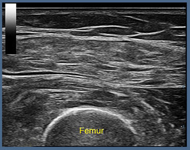
This preflight stage is a pre-treatment review using medical-grade ultrasound probe at 14 MHz frequency with 12 cm penetration and axial resolution of 0.5 mm. Conducted is a transverse scan of the quadriceps muscle. The top set of lines (labeled subcutaneous tissue). Right underneath the firm, fascia is located and between the subcutaneous fascia and the curved white bony femur outlined on the bottom is the interdigitating muscle tissues that form the four tendons of the quadriceps near the patella.


Scan #2 of 7: M-MODE ECHO-CARDIOGRAPHY (Pre Quad) 8/18/2023 1:05:39pm

Real-time scan DURING PEMF exposure
To document motion changes induced by the pulse electromagnetic pulsations, we use the motion mode setting that is used for echocardiography to demonstrate the subtlest movements in the muscle tissue inside the heart. The ammo line is seen as a white band that goes from the skin surface right down and through the curved bony portion of the femur. And the picture below this shows a steady band of horizontal lines without any motion or any change indicating that there is no contractility or motion present in the resting muscle.


Scan #3 of 7: DOPPLER/ No Flow in Box 8/18/2023 1:01:21 pm
Above the level of the femur, there's a box, which is a flow descriptor as this area is looking for arterial or venous blood flow. Notice the color bar on the top left showing directional flow where red is flow towards the probe and blue is flow away from the probe in the box. There is no evidence of color or evidence of any vessels with very low flow that may not show up that are dilated in response to the energy being put in. Hence, applying 3D Doppler Blood Flow feature of the ultrasound, we see NO blood flow or N0 Vessel dilation- indicating very low velocity that is not appearing in hemodynamic readings.
- To Be Continued - 


VISUALIZING THE TREATMENT OF INFLAMMATION
The Answers Collected from Ultrasound Imaging


Historically speaking, the most favored (and sensible) way to identify the results of any treatment is by tracking the body's immediate response to it. Controlled testing must show the patient's condition PRE and POST effects, where true data-finding is collecting the necessary EVIDENCE of its claims. The investigator can pull a significant amount of data from this form of validation testing: including stage-by-stage bodily response to future projections of possible side effects.  Modern diagnostic science looks to imaging for its safe, non-invasive yet quantifiable analyses of what’s under the skin. In this latest review, ultrasound offers a flicker-free visual (10-30 frames per second) of muscle contraction.  The “real-time” advantage of video under a 3D Doppler Ultrasound easily and clearly shows the frequency of the muscle bundle’s firing (twitching) indicating fatigue and potential pathology.


- Continued -


Scan #4 of 7: TREATMENT POWER 10/20 8/18/2023 01:05:39 pm
With the power/frequency setting of 10/20, we employed the circular transducer around the quadriceps in a 360 degree at figuration.  The motion of the muscles above this bony femur is seen to have changed from its nice, avoid appearance into something with a midline central ”waist” due to energy transfer to and within the quadriceps muscle.


Scan #5 of 7: M-MODE – PHASIC PULSATILITY NOTED 8/18/2023 01:06:56 pm
Notice the white line in the center of the switching muscle that translates into the transverse linear, and at this time, undulating or contracting muscle fibers as pointed out by the arrow at the initial pulse that reaches the muscle. Below the arrow are motion artifacts that transform the uniform, linear, horizontal lines into an unrecognized pattern of interrupted lines indicating the energy transfer and motion occurring within the muscle structure. The yellow brackets indicating pattern interruptions of muscle stasis showing visible evidence of the energy frequency induction from the PEMF.


Scan #6 of 7: DOPPLER FLOW BOX | 1.8mm VENOUS DILATION - 8/18/2023 01:11:46 pm
Measurement of a dilated vein is visually represented in this doppler flow box of the treated area between the fascia and the bone showing a dilated vein that was previously not present during pre-treatment. The vein measures 1.5 by 1.8 millimeters, and the dilatation implies blood flow of such small magnitude that it doesn't show up as a color on the registration software as the blood flow is too low. However, the fact that the veins are dilating indicates a vascular response to the energy input. The two plus marks are measurement indicators of the vein: at 1.8 mm. That's the vein that that now appeared during PEMF exposure.

Scan #7 of 7: ELASTOGRAPHY
A final (theoretical) step in this pilot study is to employ the diagnostic capacity of the elastogram feature of our ultrasound system.  In pursuit of physiological reactions to the PEMF energy induction, we may anticipate fibrosis or hardness or stiffness.  According to this scan image, we are able to capture quantitative data in the form of abnormal color which tracks with the value of the hardness or physical elastic change in the muscle fibers. The elastography scale (upper left of screen) from 100 represents very hard tissue or muscle spasm or fibrosis down to zero, indicating fat or soft tissue.  From this test, the elastogram reading shows a biometric change from 60 in hardness or spasm down to 30, representing muscle relaxation, thus adding further evidence to the PEMF effects on the condition of musculator.

CONCLUSION:  During the pre-treatment (at-rest) phase, there is no identifiable venous dilatation or blood flow. There was no muscular contractility detected by the ultra-sensitive cardiac motion mode technology.  Following a treatment at half power (10 out of 20) we can visibly and quantifiably identify that small veins are beginning to dilate and that the contractility of the tissue being energized is appearing as pulse vibrations in the form of the undulating surface line of the muscle. Measurements of change in the internal motion of the muscle fibers are outlined by the brackets (slide 5) immediately during bioenergy/electromagnetic exposure. This phase 1 test was designed to capture preliminary and evidential data of IMMEDIATE muscle/physiological reaction to energy induction therapy.  Upon further review of this quantifiable evidence and the physiological behavior of the musculature, one can extrapolate more venous dilatation and more visible blood flow activity if/when more treatment time is provided.


TECH TALK:
"Choosing The Right Scanner for My Clinical Research Project":

For this pilot study, recognized diagnostic imaging specialist Dr. Robert Bard selected the latest feature-rich MINDRAY ultrasound system: the RESONA 7. "One of my passions is learning about the latest advancements in ultrasound imaging... and thanks to a recent industry showcase, the Resona 7 stood out to me as a well-designed and highly flexible scanner that can handle the many challenges of an exploratory research project."

MANUFACTURER'S OVERVIEW:
The Mindray Resona 7 platform begins with proprietary and industry-first virtual beam-formation technology, ZONE Sonography® Technology+. The acquisition of larger and more robust acoustic data sets coupled with proprietary, industry-first virtual beamforming methods and innovative signal processing techniques translates into a number of unique imaging advantages. These include extremely fast image processing and display for noticeably improved temporal resolution; exceptional image uniformity throughout the field of view with improved spatial and contrast resolution. ZST+ also has the ability to generate novel and exclusive advanced diagnostic applications while improving the performance of established imaging technologies. Virtual beam formation will continue to shape the future of ultrasound imaging in impactful ways, allowing for more powerful technology, improved workflow tools, and the inclusion of more AI-based advancements. ZST+ has paved the way for the future of ultrasound.

Mindray’s High Fame Rate Sound Touch Elastography utilizes the advantages of ZST+ with smoother transition times and faster refresh rates for more consistent acquisition and assessment during live 2D shear wave imaging allowing for more rapid, reliable, and reproducible quantitative results.  

Advantages include:
- Patented ultra-wide beam tracking technology improves penetration, especially for stiff tumors, fatty livers, and obese patients
- Rapid acquisition with high reliability for improved efficiency and throughput
- Consistent results with high reproducibility
- Four quality measures for improved inter-operator variability and diagnostic confidence: motion stability, tissue homogeneity, reliability map, and reliability overlay visualization
- Continuous-fire STQ acquisition methods for more results in less time, enhancing the patient experience
- Combination STE and STQ mode for continuous-fire point measurement with the added advantage of quality maps 

For complete information on this technology, visit: www.MindrayNorthAmerica.com


* This clinical performance test was conducted by: Dr. Robert L. Bard of the AngioResearch Foundation and the Integrative Health Research Center for HealthTech Reporter.  Our clinical diagnostic testing team gives special thanks to MINDRAY, NA and AURAWELL PEMF for the equipment donated in the execution of this performance study- without whose generous support this study could not have been made possible.

All images, analytical content and narrative graphics presented in this report are copyright © 2023- Bard Diagnostic Imaging. All Rights Reserved.


Friday, September 1, 2023

IMAGE GUIDED APPROACH TO THE TREATMENT OF PROSTATE HYPERPLASIA

Written by: Robert L. Bard, MD

Procedurally, the best way to study an enlarged hyperplastic prostate gland is as a low grade inflammatory process, similar to women with fibrocystic breasts considered to be breast inflammation. [1] This does not need to be treated with antibiotics since this inflammation is often aligned with a chronic disease and requires a long-term treatment protocol. One example of this therapy is using a pulse electromagnetic applicator (PEMF) which can be applied over the groin area or under the pelvis (even in the car seat). For treatment applications, this protocol is recognized to be safe, painless, comfortable, and user-friendly on a long-term basis. 

Reducing low grade inflammation can be managed with relatively low energy treatment over an extended yet controlled period of time.  Daily application of energy therapy inducers to the prostate and bladder area has been proven to reduce bladder urge, the need to urinate and improve the urine flow out of the enlarged prostate. It also decreases the chance of incontinence because the bladder that is more easily emptied is less likely to overflow in an uncontrolled manner.


PEFORMANCE REPORT 1: The objective of this limited study was 2 fold: To assess for any unexpected side effects and demonstrate physical changes in the treated gland with at home PEMF treatment.   

Results: No adverse effects were reported in 3/3 subjects. 75% of the group had baseline and 30 day followup volumetric ultrasound with 3D high resolution probes and demonstrated variable volume loss. From 2 years of experience with the given PEMF device, it is understood that these technologies are not uniformly adaptable to all patient types with the uniform probe positioning for this study and limited length of time for the procedure. A full Phase 1 study with standard urologic measurements and ultrasound measurements of: 

1‐blood flow perfusion of inflammatory microcirculation
2‐elastography of physical characteristics‐scarring or fibrosis
3‐presence of macrocalculi or change in microcalculi
4‐presence of bladder pathology (stone, tumors, postvoid residual, hypertrophy) 


CASE STUDY 1: Caucasian male, 69 years old 

SONOGRAM: RE+ right psa elevated  Comparison 7/5/23 1 month PEMF 

TIME OUT: risks/benefits/possibility of false positives discussed 

Prostate, bladder and pelvic paraprostatic  spectral Doppler vascular study was performed using  transrectal 4D  real  time b mode examination, power Doppler, spectral flows and 3D workstation analysis. This report sequence was limited to volumetric changes based on non invasive therapies.  Dre=+/‐ left  

PROSTATE VOLUME:
Base line: Previous pre‐treatment prostate vol: 104cc
After Exposure Effect: Current post treatment prostate vol: 92cc 

Vascular area: r lateral 9x7 prev 9x7 mm.  VI=1%  unchanged Vessel Index is a quantitative measure of inflammatory or malignant vessels.  Note the absence of increased vascularity in the 9x7 mm focus signifies no evidence of abnormally induced vessel related adverse effects over the one month study period.

OVERVIEW OF ENERGY INDUCTION: Both benign enlargement and increased incidence of cancer are associated with micro inflammation.  PEMF therapy may result in a reduction in kidney obstruction, a decrease in urinary symptoms and a reduced incidence in cancer as a benefit of routine use may be forthcoming.


PSA READINGS vs IMAGING

In 1930, the PSA was developed by the University of Arizona as a rape test. It was accurate for finding and identifying semen in rape victims but it was never made to be diagnostic for prostate cancer. The 2004 Journal of Urology stated the PSA is 2% accurate in diagnosing prostate cancer definitively, which means that the digital rectal exam is at least three times more accurate than the PSA blood test in finding clinical prostate cancer. Also, in benign diseases such as prostatitis or benign enlargement with older age, the PSA level automatically rises in most patients. Therefore, the PSA lifespan has ended, much like the x-ray has ended in the use of prostate disease because we use CT MRI and ultrasound.  CT and MRI is being replaced by the imaging community is now trending to substitute 3D Doppler and elastography to scan the prostate for prostate cancer aggression and capsule integrity worldwide [4].


PEMF FOR PROSTATE HEALTH
Current research reports show that PEMF has been applied clinically in prostate therapeutic treatments [2,3]. Under the philosophy of inflammation reduction, our pilot study was designed to test PEMF performance and the patient's physiological reaction before and after treatment as we are evaluating the treatment effect in real time. 

Because we are able to calibrate the strength of the prostate bioenergy entering the body by monitoring the autonomic nervous system we were able to fine tune the therapeutic process.  So far we are having measurable success in positively addressing symptoms and (hopefully) will have proven over time a long term goal that reducing prostate inflammation also reduces the incidence of clinical prostate cancer.

It is important to note that low grade inflammation is oftentimes a precursor or potentially mixed with cancers. The possibility of reducing prostate cancer risk by using post bioenergy noninvasive protocols is something that makes clinical sense and has been widely used in other countries [5,6]. Upon the strategic design of our current pilot study, we hope to expand the scientific findings of this protocol to lead to greater use of non-invasive pulsed energy solutions for prostate and allied health problems.


References: 

1) "Enlarged Prostate" - PennMedicine.org https://www.pennmedicine.org/for-patients-and-visitors/patient-information/conditions-treated-a-to-z/enlarged-prostate#

2) "Effect of Pulsed Electromagnetic Field Therapy on Prostate Volume and Vascularity in the Treatment of Benign Prostatic Hyperplasia: A Pilot Study in a Canine Model" | National Institutes of Health.- PMID: 24913937- Raffaella Leoci,* Giulio Aiudi, Fabio Silvestre, et al.  https://www.ncbi.nlm.nih.gov/pmc/articles/PMC4145661/

3) "Therapeutic use of pulsed electromagnetic field therapy reduces prostate volume and lower urinary tract symptoms in benign prostatic hyperplasia"- PMID: 32090492 |  Marta Tenuta, Maria G Tarsitano, Paola Mazzotta et al. https://pubmed.ncbi.nlm.nih.gov/32090492/

4) "Ultrasound & The Prostate": RadiologyInfo.org |  https://www.radiologyinfo.org/en/info/us-prostate#:~:text=Ultrasound%20of%20the%20prostate%20uses,whether%20the%20gland%20is%20enlarged.

5) "Prostate cancer ultrasound treatment as effective as surgery or radiotherapy", by Kate Wighton (04 July 2018). Imperial College London. https://www.imperial.ac.uk/news/187086/prostate-cancer-ultrasound-treatment-effective-surgery/

6) "Mechanisms and therapeutic effectiveness of pulsed electromagnetic field therapy in oncology"- 2016 Nov 5, | PMID: 27748048 | Maria VadalĂ , Julio Cesar Morales‐Medina, Et. Al. - https://www.ncbi.nlm.nih.gov/pmc/articles/PMC5119968/


ASSOCIATE CONTRIBUTOR:

Dr. JERRY DREESSEN is both Trauma Team Qualified and Hospital Qualified treating chiropractor in Mountlake Terrace, WA. He is the Executive Director for the Association of PEMF Professionals- the largest professional PEMF organization in the United States. AOPP attracts the most principled and accomplished PEMF user- setting standards by providing certification for all PEMF users, as well as Continuing Education (CEU's) for all members to maintain best practices and techniques. (www.backtoaction.com)


SPECIAL THANKS
The news feature proudly gives thanks to the AOPP (Association of PEMF Professionals) and Patrick Ziemer of Magnawave Inc. and Aura Wellness PEMF for the technical information provided for interviews and unending support to support our clinical study and educational programs about PEMF technology for chronic disorders and supportive testimonials in alternative therapeutics.   Additional special thanks to Dr. Robert Bard and  "Cousin Sal" Banchitta and Russ Allen (Wellness NOW!) for their proactive leadership in support of men's health in this review under the "Get Checked NOW!" advocacy program. 

Beyond the Website: Auditing the Digital Health of a Business

  A Technology Report & Review with Dave Olsen of Hassle Free Web INTRODUCTION: Your Website Is Only One Part of Your Digital Business F...